Contents
Overview
Learn how to predict which glycans are likely to bind to a given protein.
To do this, we will use the Gly-Spec Webtool, which uses the 3D structure of a glycan-binding protein in complex with a carbohydrate fragment that is a minimal binding determinant (MBD) for that protein. The webtool finds glycans that contain the MBD within their structure, and predicts if they will bind to the protein.
Example System
This scenario can be followed with a co-complex of your choice, but if you don’t have one, the following files have what you need to follow along:
The zip file contains a pdb file of the UDA lectin (Urtica dioica agglutinin), along with a glycan fragment that has been trimmed down to only the MBD. It also contains a simple text file which is a list of the glycan IDs of several glycans that are known to bind to UDA.
Background
The Gly-Spec tool was originally named the Grafting tool, and it was developed by Oliver Grant. The term Gly-Spec refers to the specificity of glycans.
What the tool does:
The Gly-Spec tool takes a protein in complex with a glycan fragment representing the MBD and scans a user-selected glycan array, or the human glycome, for glycans that contain the MBD.
The Gly-Spec tool then tries to build each of the glycans it found into the binding site of the protein, by growing out the larger glycan from the co-complexed MBD. If the attached “branches” have any atomic overlaps with the protein binding site the webtool attempts to resolve them by “wiggling” the interglycosidic linkages within limits that can be set by the user. In cases where the adjustments do not resolve the overlap, the tool predicts that the glycans will not bind. In cases where all overlaps are resolved by wiggling, the tool predicts the glycan will bind.
The Gly-Spec tool also considers whether the linkers used on a CFG glycan array might prevent binding of a glycan that otherwise would bind, and reports this to the user.
Finally, the Gly-Spec tool produces a report that lists predicted binders and non-binders, compares to any provided list of binders, and gives the user the opportunity to download the 3D structure files for the glycans that were screened. This includes both the predicted binders and non-binders. These can be viewed in programs like VMD, to visually demonstrate the structure of binders in complex with your protein, or to assess the particular protein residues or structural features that prevent certain glycans from binding.
Required Input
The tool requires a pdb file of a protein and a glycan fragment in complex. If you don’t have a co-complex, and just want to learn, see the Example System section above.
Optionally, we can also upload a text file containing a list of the glycan ids of known binders. Most often you want to, because this allows the tool to estimate its accuracy. Known binders are usually determined as the result of experimental screening data.
Protocol
The Gly-Spec tool can be found here.
Preparing Input Files
Co-Complex
Prepare your input so that your PDB file contains an glycan fragment in complex with a protein. You want to be sure that:
- Your protein is contained in the ATOM section
- Your glycan is contained in the HETATM section
- Your glycan portion only contains the atoms that are in the MBD, however:
- It is ok to guess, and use the tool to assess your guess
- Also try to make sure that the glycan does contain the whole MBD
- Be sure to remove any duplicate copies of the oligosaccharide
- Make sure that all residue numbers are unique
Known Binders
Optionally, you can also provide a list of known binders for comparison. It is ok not to provide these, but they can be used to assess the accuracy of the predictions made by the tool.
This file should be a simple text file that contains a list of glycan ids, one per line, identifying glycans that were shown to bind in experimental data. The file in our example system looks like this:
216
212
51
217
211
352
50
213
347
Note: Be certain that your numbers match those of the Library that you choose below!
Upload Input
Use the Choose File button to upload your co-complex:
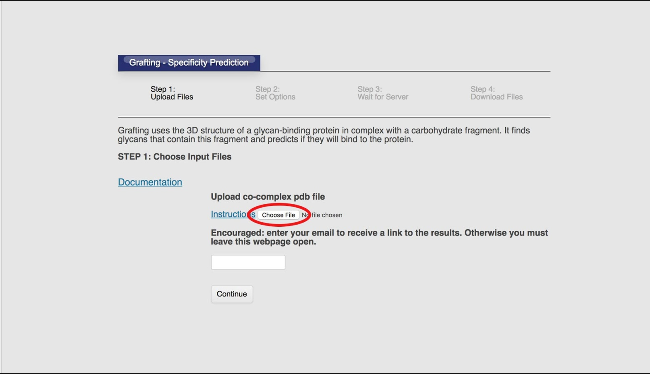
Optionally, enter your email address into the the text field. This is highly encouraged, because if you don’t, you will need to leave the page open until the tool is finished. Entering your email address allows the tool to email you when your job is complete with a link to the results.
Click continue.
Set Options
In this step, we decide which glycan array to scan, or possibly the known human glycome, set the maximum torsion range (in degrees), and upload any known binders.
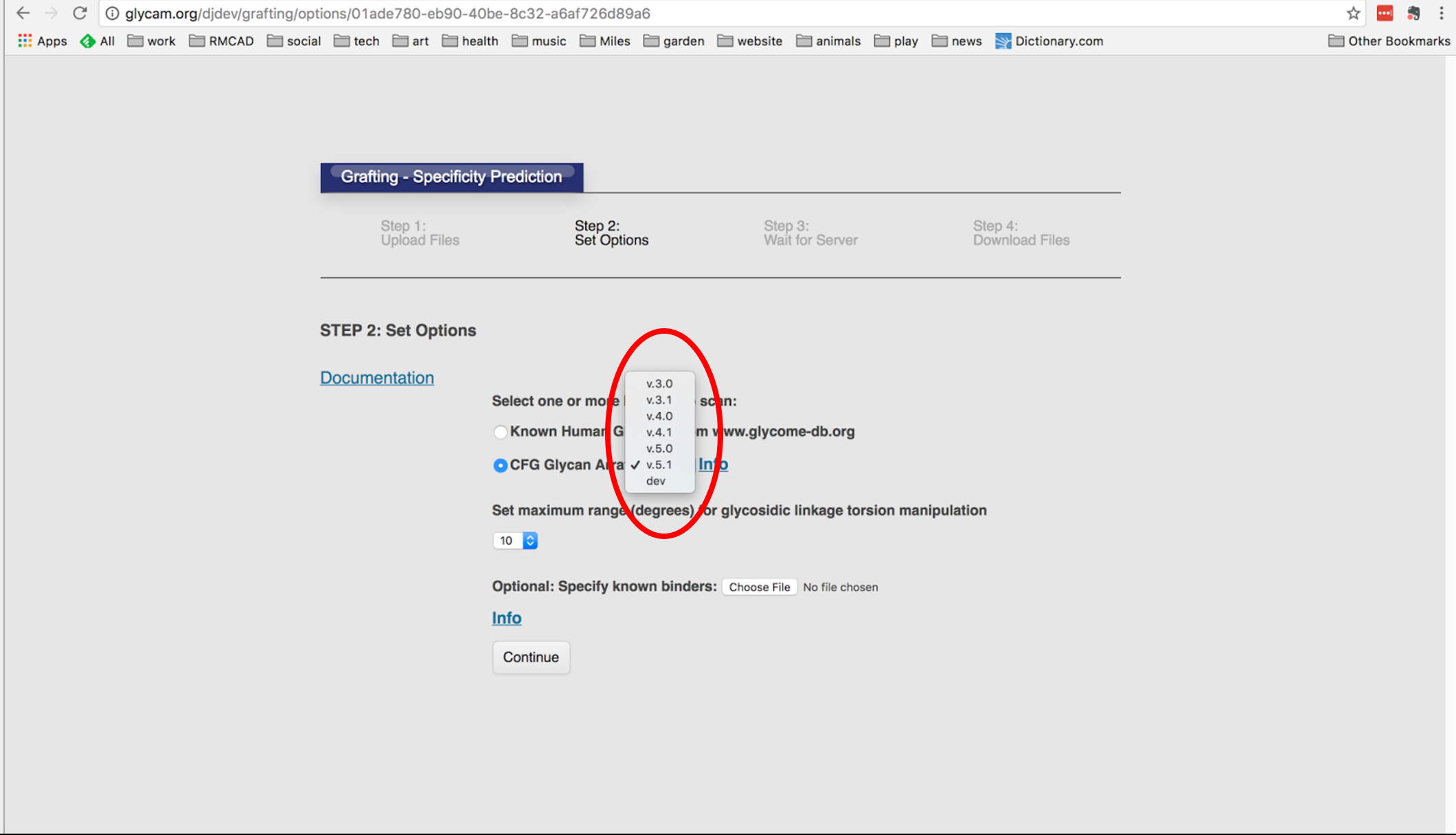
Selecting A Library
Either select the Known Human Glycome, or select one of the CFG Glycan Array versions from the picklist:

or select a CFG library version to scan:
Set a maximum range (in degrees) for glycosidic linkage torsion manipulation:
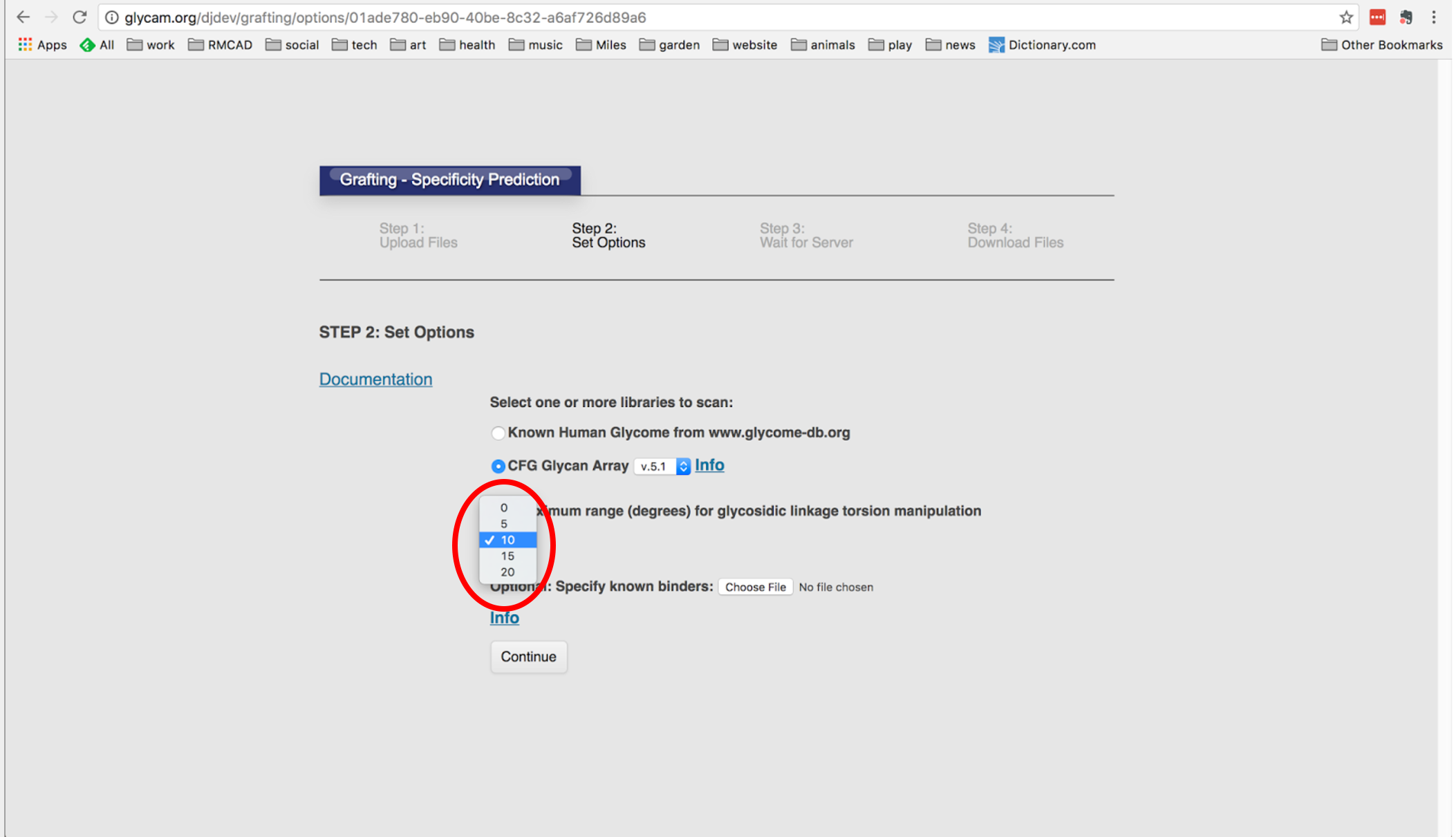
Optionally, upload a text file of known binders:

Click Continue.
Wait For Processing
The next step involves waiting for the tool to create output. The range of wait times is fairly large: some simple jobs can finish in a few minutes, while more complex systems can take hours.
If you entered your email address, you can close the web page because you will receive an email when your job is completed, along with a link to the page that allows you to see your results.
Interpret The Results
The tool provides you with a report summarizing the results, along with a zip file containing the 3D structures of all of the scanned glycans, separated into predicted binders and predicted non-binders.
The report lists details about each scanned glycan:
- Array ID: Glycans identified sequentially.
- Exp Bind?: “Y” if experiment array data showed a bind, “N” otherwise
- Pred Bind?: “Y” if the grafting tool predicts a bind, “N” otherwise
- Exp & Pred Agree?: Good if both Exp and Pred are “Y” or “N”, Bad if they differ
- Spr Eff?: “Y” if there is a spacer effect, “N” otherwise
- Spr ID: “N/A” if no spacer effect. ID of spacer otherwise.
- Glycan Structure: text notation of the glycan, e.g. DGlcpNAcb1-2DManpb1OH
The report also provides a summary that details:
Text summary:
- How many non-binders were correctly predicted
- How many binders were correctly predicted
- How many cases were improved by modelling the linker
- How many cases were less accurate when modeling the linker
- List of any experimental binders which did not contain the uploaded sequence
- Job ID
- The complex provided by the user
- The carbohydrate detected from that complex
- Which glycan library was scanned
- The email address provided by the user
Annotated Output
The 3D structures are provided in the form of PDB files. These can be viewed in modeling applications, such as VMD.
Additional Info
In case someone wants additional information about this or that.
Alternate Protocols
We are still developing the alternate protocols.
Troubleshooting
Tips for the user.
